Ultrasound Scan- Step By Step Procedure?
Body

When Is An Ultrasound Recommended?
There are several different types of ultrasound scans. For example, an ultrasound test may be external, internal, or endoscopic. The type of scans depends on the specific body parts that need an examination to detect any abnormality.
- Abdominal Ultrasound examines the internal organs of the abdomen, such as the liver, gallbladder, pancreas, and spleen.
- Obstetric or pregnancy Ultrasound is a scan to assess the growth of the fetus.
- Female pelvis ultrasound uses a transducer in the vagina or externally to detect any problem in the pelvis, uterus, cervix, fallopian tubes, and ovaries.
- Breast ultrasound is used to assess breast symptoms like lumps and cancer.
- Renal Ultrasound scans the urinary tract, including the kidneys and bladder.
- Transrectal Ultrasound investigates problems related to the prostate gland.
- Doppler Ultrasound monitors blood flow in the major arteries and veins.
- An echocardiogram examines the heart.
- 3D Ultrasound shows a three-dimensional picture of the internal organs
- 4D Ultrasound creates a three-dimensional picture in motion for better diagnosis.
- Internal Ultrasound scans involve placing an ultrasound probe into the vagina or rectum.
- Endoscopic Ultrasound or Echo-endoscopy is a procedure in which a probe is inserted into a hollow organ. Ultrasound produces images of the internal organs in the chest and abdomen. It is used in the upper digestive and respiratory systems.
What Does An Ultrasound Scan Reveal?
Physicians recommend ultrasound tests for screening and diagnosis.
- To examine the thyroid gland, the breast, the prostate, and the liver.
- To detect the muscles, tendons, and ligaments and investigate sprains, strains, trapped nerves, and muscle tears.
- To check if there are lumps in the breast and recommend further tests to distinguish between fluid-filled cysts and solid lumps.
- Monitor blood flow and identify blood clots, bulging, and narrowing arteries.
- To guide the doctors for treatments, Ultrasound shows the correct place for an injection or a biopsy needle.
- To monitor the growing fetus during pregnancy and check the baby's health.
- To diagnose gallbladder disease.
- To check the thyroid gland.
- To evaluate genital and prostate problems.
- To assess joint inflammation.
- To evaluate metabolic bone disease.
- Ultrasound has proven its benefits in pregnancy, including assessing gestational age, monitoring the fetus's progress, and screening for any complications. This scan is usually done during 8 to 13 weeks and 18 to 20 weeks.
How Is An Ultrasound Scan Done?
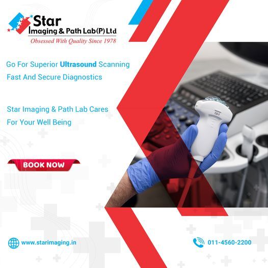
An ultrasound test is performed using a handheld transducer connected to a computer. High-frequency sound waves are sent, which bounce back and are converted into electrical impulses that come up on a monitor.
The patient is asked to lie on the back or side. Ultrasound gel is applied on the skin where the scan will take place. The radiologist moves the transducer on the gel. Usually, he presses a little hard on the skin to get the pictures more accurately.
During a transvaginal ultrasound scan, the patient is asked to empty the bladder and undress from the waist down. A sheet is provided to cover up. It is lubricated with gel and inserted into the vagina. The radiologist gently moves it around to capture pictures. Women can request a female radiologist for this type of Ultrasound.
A transrectal ultrasound is done when a narrow transducer, coated in gel, is inserted into the rectum to take images of the prostate and its surrounding tissues. This does not involve any pain.
Search ‘ultrasound scan near me’ to find out about the diagnostic centers around your area. Our Ultrasound scan center is well equipped with the latest facilities to serve you better. Our professionals and health care assistants are experienced and trained to help you in every possible way. We provide accurate results within the stipulated time. To know further, contact us.


Comments